Blog Layout

Focus on Shin Splints

October 14, 2024

What is it?

Shin splints, are a painful condition of the lower leg, also known as Medial Tibial Stress Syndrome, it is an overuse injury that causes pain along the inside of the tibia or shin bone. It is a common condition in runners, hikers and soldiers who march long distances.

What are the symptoms?

Shin splints are typified by persistent leg pain, usually the inside of the shin, halfway down the lower leg. The pain might be felt during exercise or directly after. Some people experience a dull ache over their shin that lasts for quite a while after exercise stops, while for others the pain may be sharp and fade quickly. The pain is often progressive, becoming worse with shorter distances. Eventually, shin splints can severely impact activity levels as the pain becomes too severe to continue exercising. 

Shin splints can be extremely painful and very disruptive to activity levels. As the pain usually starts gradually and progresses many people find themselves unable to continue training, shin splints may also progress to stress fractures if not diagnosed early and managed effectively.

Shin splints are typified by persistent leg pain, usually the inside of the shin, halfway down the lower leg. The pain might be felt during exercise or directly after. Some people experience a dull ache over their shin that lasts for quite a while after exercise stops, while for others the pain may be sharp and fade quickly. The pain is often progressive, becoming worse with shorter distances. Eventually, shin splints can severely impact activity levels as the pain becomes too severe to continue exercising. 

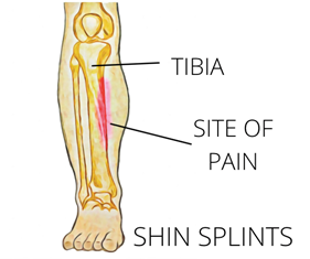
A diagram of a shin splint showing the location of pain

How does it happen?


Shin splints are predominantly seen in runners who increase their distances quickly, often while training for an event. Activities that require repetitive weight-bearing of any kind, such as marching or high impact sports have also been shown to cause shin splints. Although the pathology of shin splints is unclear, studies have been able to identify certain risk factors that may predispose someone to shin splints. These include;

  • An abrupt increase in activity level
  • Improper footwear and support
  • Higher BMI
  • Training on hard or uneven surfaces
  • Tight calf muscles
  • Flat feet
  • Increased external rotation range of the hips
  • Females are more likely to develop shin splints than males.
  • Prior history of shin splints
  • Wearing or having worn orthotics


How can physiotherapy help?


The first step for your physiotherapist will be to address any contributing factors and help to adapt your training program to a level that is optimum for you. A period of relative rest may be recommended along with a targeted strengthening and stretching program for any tight or weak muscles. Switching to low-impact activities such as swimming, cycling and yoga may also help to maintain fitness during recovery. Your running technique will be analyzed and any training errors may be corrected. When getting back into your training routine, it is usually recommended that distances are not increased by more than 10% per week as this allows the tissues of the body to react to the increased demands and adapt accordingly.


None of the information in this newsletter is a replacement for proper medical advice. Always see a medical professional for advice on your injury. 

Shin splints can be extremely painful and very disruptive to activity levels. As the pain usually starts gradually and progresses many people find themselves unable to continue training, shin splints may also progress to stress fractures if not diagnosed early and managed effectively. 


How does it happen?


Shin splints are predominantly seen in runners who increase their distances quickly, often while training for an event. Activities that require repetitive weight-bearing of any kind, such as marching or high impact sports have also been shown to cause shin splints. Although the pathology of shin splints is unclear, studies have been able to identify certain risk factors that may predispose someone to shin splints. These include;

  • An abrupt increase in activity level
  • Improper footwear and support
  • Higher BMI
  • Training on hard or uneven surfaces
  • Tight calf muscles
  • Flat feet
  • Increased external rotation range of the hips
  • Females are more likely to develop shin splints than males.
  • Prior history of shin splints
  • Wearing or having worn orthotics


How can physiotherapy help?


The first step for your physiotherapist will be to address any contributing factors and help to adapt your training program to a level that is optimum for you. A period of relative rest may be recommended along with a targeted strengthening and stretching program for any tight or weak muscles. Switching to low-impact activities such as swimming, cycling and yoga may also help to maintain fitness during recovery. Your running technique will be analyzed and any training errors may be corrected. When getting back into your training routine, it is usually recommended that distances are not increased by more than 10% per week as this allows the tissues of the body to react to the increased demands and adapt accordingly.


None of the information in this newsletter is a replacement for proper medical advice. Always see a medical professional for advice on your injury. 
September 9, 2024
Most tissues in the body have healed completely in six to 12 weeks following an injury, however, many people have severe pain that lasts much longer than this. We know that the intensity of the pain you feel is not always associated with a similar amount of damage. In some cases, there can be a severe amount of pain with almost no detectable damage. With this in mind, we explore some reasons why your pain might not be getting better, long after the tissues have healed. You’re afraid of the pain. Pain can mean many different things, for some of us pain can affect our ability to work or can be a symptom of a serious disease. What you believe about your pain can either amplify or reduce the symptoms you experience. If you feel that every time you experience pain you are causing more damage, you will naturally pay more attention to this and your nervous system will amplify the signals in an attempt to keep you safe. If you understand the cause of your pain and know that while there is discomfort, you are not in danger of causing more damage, often the pain will feel less severe. This is one of the benefits of seeing a physiotherapist after your injury as they can help you to understand your pain, giving you more control over your recovery. You started moving differently after the injury. Immediately after an injury, it’s natural to change the way you move to avoid painful movements. After a while, these changed movement patterns can become maladaptive and actually begin to cause pain and discomfort on their own due to the altered stress patterns placed on your body. Correcting these adaptive movement patterns can often go a long way in reducing pain after an injury. You might not have noticed these changes and might need a physiotherapist to identify and help you to return to your usual movement pattern. You have lost muscle strength since the injury. While a certain amount of rest following an injury is always helpful, if we stop moving altogether, our muscles can lose strength. This can mean that our posture changes, we fatigue easier during our usual activities and that we are more susceptible to further injury. Less movement also means we actually focus on the pain more when it does happen. Physiotherapists are able to advise you on the right types and amounts of excercise for you in the period following your injury. The pain has affected your lifestyle. When pain affects your ability to sleep, work and even concentrate, it’s not surprising that this can have a negative affect on your overall wellbeing and mental health. This can create a negative cycle of anxitey and depression that perpetuates and increases the experience of pain. If your pain is really getting you down, speaking to a mental health professional can actually be a valuable part of your physical recovery
September 9, 2024
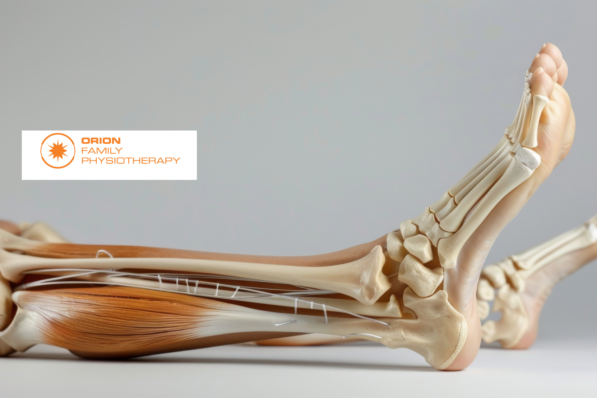
Tendons are found all over the body and while you may know a little about them, you might be surprised to learn a few of these facts.
September 9, 2024
10 Facts About Tendons 
May 24, 2024
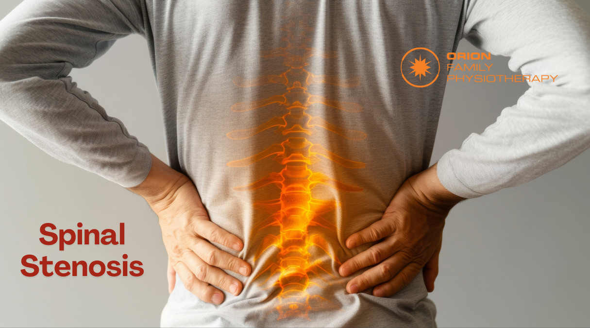
What is spinal stenosis? The spinal cord, nerves and arteries are housed by the spine, which acts as a hard electrical casing to support and protect these vulnerable structures. The spine has a hollow column that allows the spinal cord to run from the brain to the rest of the body. At each spinal segment, nerves exit the spine and supply the tissues of the body. There is also an intricate network of small veins and arteries that provide blood to the spinal cord and vertebrae, providing them with the nutrients needed to operate. Spinal stenosis is characterized by a narrowing of the spaces that house the spinal cord, nerves and blood supply. A variety of factors can cause spinal stenosis, however overwhelmingly it is caused by degenerative changes to the spine as we age. Many people over the age of 60 will have spinal stenosis; however, not all will have pain. Clinically, spinal stenosis is used to describe the painful symptoms of this condition rather than just the narrowing itself.
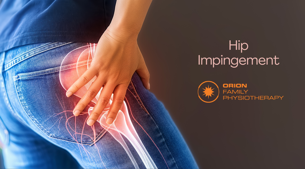
Hip Impingement - Springfield Central, QLD - Orion Family Physiotherapy
May 23, 2024
What is it? When the two surfaces of the hip joint move over each other, they usually move freely without any friction. If there is an alteration to either the socket part of the joint (the acetabulum) or the ball (the head of the femur), irritation may occur as the two surfaces move over each other. This is known as Femoroacetabular impingement, a common disorder of the hip, characterized by pain and stiffness. Femoroacetabular impingement can be classified as cam, pincer or mixed. A cam FAI occurs when the femoral head junction is flattened or a small bump is present. Pincer type of impingement occurs when the acetabular rim extends slightly, causing the femur to be impacted. Cam impingement is more common in men while pincer impingement is more common in women. However, most cases of FAI (about 85%) are mixed, meaning they both have cam and pincer types of impingement.
Man With Poor Balance
By Sensis Master March 26, 2024
Ankle sprains are one of the most common sporting injuries and most people have experienced one at least once in their lifetime. While they are common, this doesn’t lessen their negative impacts. Surprisingly, having poor balance might be increasing your risk of ankle sprains. Here we discuss a few facts about balance and what you can do to reduce your risk of ankle injuries.
Muscular Trigger Points
By Sensis Master March 26, 2024
What Are They? Muscular trigger points are better known to most of us as muscle knots and can feel like painful, hard lumps located inside muscles. These knots can both be painful to touch and refer pain in surrounding areas. It is thought that trigger points form when a portion of muscle contracts abnormally, compressing the blood supply to this area, which, in turn, causes this part of the muscle to become extra sensitive. Trigger points are a common source of pain around the neck, shoulders, hips and lower back.
Injury Healing
By Sensis Master February 6, 2024
When injury strikes, the first thing that most of us want to know is ‘how long will this take to heal?’ Unfortunately, the answer to this can be complicated and requires at least a little understanding of how the different tissues of the body heal. Each of the tissues of the body, including muscles, tendons, ligaments and bone, heal at different speeds and each individual will have some variation on those times as a result of their individual health history and circumstances.
Golfer’s Elbow
By Sensis Master December 12, 2023
Golfer’s Elbow (Medial Epicondyle Tendinopathy) Golfer’s elbow is tendinous overload injury of the tendon on the inside of the elbow, usually due to overuse. As its name implies, it is a condition common in golfers. However, as with all sporting injuries, this condition can affect anyone. Golfer’s elbow is similar to Tennis elbow, occurring on the inside of the elbow rather than the outside. What are the symptoms? Typically, someone suffering from this condition will experience pain on the inside of the elbow, forearm and possibly extending down to the hand. The pain will be worst with activities that require gripping of the hand and movements of the wrist. Less common is the experience of pins and needles in the hand. How does it happen? The exact cause of this condition is unknown; however, it is generally thought to occur when the forces transmitted through the tendon become too great. This can be due to increased demands on the tendon or reduced quality of the tendon tissues. As the tendon is attached to muscles that bend the wrist and provide grip strength, activities such as golf, rock climbing or manual work that involve gripping objects can easily create forces that damage the tendon. Conversely, factors such as poor blood supply or simply the normal processes of aging can reduce the quality of the tendon. If the tissue is not functioning well, then even simple but repetitive movements in an office job can cause Golfer’s elbow. There are a few other known contributing factors for Golfer’s elbow, such as poor posture, neck dysfunction, a recent change in activity and a history of trauma, such as a fall onto an outstretched hand. What is the treatment? Golfer’s elbow usually develops slowly, and healing can be a long process. The first step to effective treatment is accurate diagnosis, as many other conditions have similar symptoms and need to be excluded first by a medical professional. Once a diagnosis of golfer’s elbow has been confirmed, treatment is aimed at allowing tissues to heal and regenerate. This will require a certain level of rest, and changes to the forces affecting the tissues, sometimes through bracing or taping. Specific exercises have been shown to assist tissues in coping with and responding to load; these are called “eccentric” exercises. Other treatments include increasing blood flow to the area to promote healing. In chronic and severe cases, injections of corticosteroids are used, and in severe cases surgery may be undertaken. The information in this newsletter is not a replacement for proper medical advice. Always see a medical professional for assessment of your condition.
Show More
Share by: